Parts of the brain including the dura mater William Say
Product images of Parts of the brain including the dura mater


Sizing information
| Overall size (inc frame) | x cm ( x in) |
| Depth | cm (in) |
| Artwork | x cm ( x in) |
| Border (mount) |
cm
top/bottom
(in)
cm left/right (in) |
| The paper size of our wall art shipped from the US is sized to the nearest inch. | |
 zoom
zoom
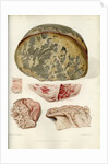
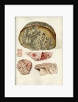
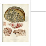
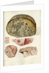
Parts of the brain including the dura mater
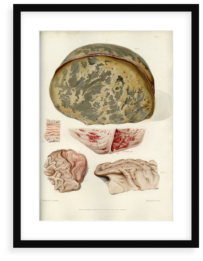
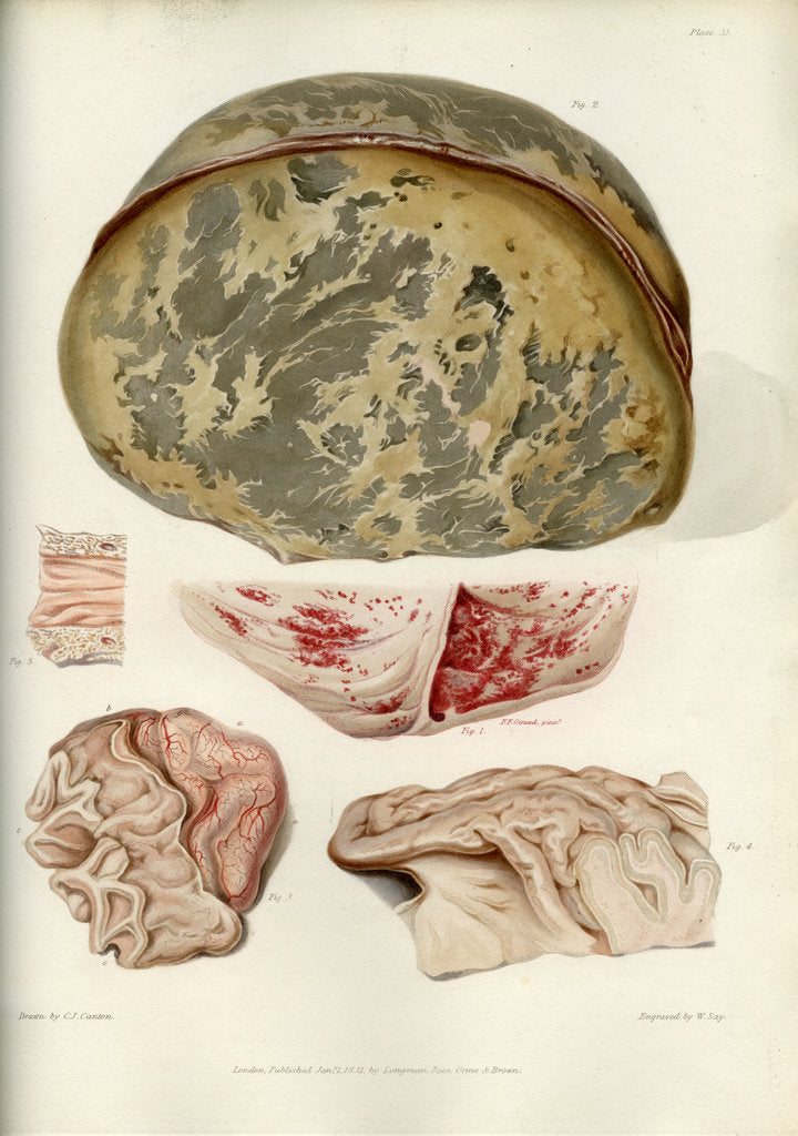
Illustration of five parts of the brain, each illustrating a disease. Fig.1 û central image. A portion of the dura mater taken from a woman who suffered from symptoms of cerebral pressure. Fig.2 û top image. Ossification of the dura mater. This was taken from a case of chronic hydrocephalus. It shows an extensive deposit of bony matter in thin fibres across the substance of the membrane. Fig.3 û bottom left image. 'A portion of the brain of a child who died with serous effusion into the cellular tissue of the pia mater and into the ventricles.' Fig.4 û bottom right image. A portion of the middle lobe of the cerebrum. Specimen from an old woman who suffered 'under symptoms of general paralysis and imbecility.' Fig.5 û central left image. A small portion of the theca of the spine taken from an elderly woman who 'laboured under an obstinate form of spasmodic wry neck.' Plate 31 from Bright's Medical Reports: Diseases of the brain and Nervous System part 2 by Richard Bright (London, Longman, Rees... 1831). Inscribed: 'Plate 31.Drawn by C. J. Canton delin. London Published Jan 1st. 1831. by Longman, Rees, Orme & Brown. Engraved by W. Say'
Original: engraving. 1831
- Image reference: RS-10318
- The Royal Society
Discover more
More by the artist William Say.
Explore the collection Science and anatomy prints and drawings.
Our framed prints
Every framed picture is created by hand in our workshop by specialist framers.
Black, white, brown, silver, gold or natural frames available, supplied ready to hang.
All our frames have a smooth satin finish, and measure 20mm (front face) by 23mm (depth from wall).
Read more about our framed art prints.
Manufactured in the UK
All products are printed in the UK, using the latest digital presses and a giclée printmaking process.
We only use premium branded inks, and colours are independently verified to last between 100 and 200 years.
Delivery and returns
We print everything to order so delivery times may vary but all framed pictures are despatched within 5-7 days via courier or recorded mail.
Delivery to the UK is £10 for a single framed print.
We will happily replace your order if everything isn’t 100% perfect.