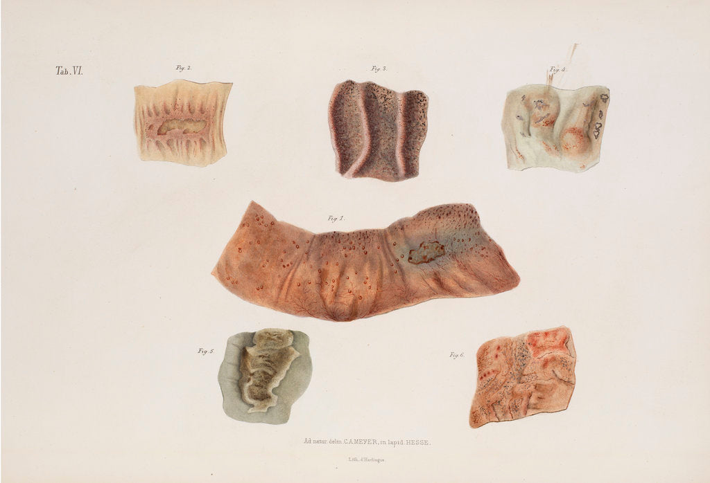
Alterations caused by cholera in the intestinal mucous membrane d'Harlingue
Product images of Alterations caused by cholera in the intestinal mucous membrane


 zoom
zoom
Alterations caused by cholera in the intestinal mucous membrane
Anatomical study showing the alterations caused by cholera in the intestinal mucous membrane during various stages of the disease. Fragment of the ileum in the typhoid stage of the disease (figure 1), choleric ulceration of a Peyer's patch (figure 2) and the blistering of the villi of the ileum (figure 3). The effect of diphtheric cholera on the large intestine demonstrating local hyperaemia and swelling (figures 4 and 6) and the effect of simple cholera on the mucous membrane of the large intestine (figure 5). Plate VI from Anatomie Pathologique du Cholera Morbus by Nikolay Pirogov (St Petersburg, 1849).
Original: lithograph . 1849
- Image reference: RS-9943
- The Royal Society
Discover more
More by the artist d'Harlingue.
Our prints
We use a 240gsm fine art paper and premium branded inks to create the perfect reproduction.
Our expertise and use of high-quality materials means that our print colours are independently verified to last between 100 and 200 years.
Read more about our fine art prints.
Manufactured in the UK
All products are printed in the UK, using the latest digital presses and a giclée printmaking process.
We only use premium branded inks, and colours are independently verified to last between 100 and 200 years.
Delivery and returns
We print everything to order so delivery times may vary but all unframed prints are despatched within 2-4 days via courier or recorded mail.
Delivery to the UK is £5 for an unframed print of any size.
We will happily replace your order if everything isn’t 100% perfect.


