'Tumour of the pons Varolii' William Say

Product images of 'Tumour of the pons Varolii'


Sizing information
| Overall size (inc frame) | x cm ( x in) |
| Depth | cm (in) |
| Artwork | x cm ( x in) |
| Border (mount) |
cm
top/bottom
(in)
cm left/right (in) |
| The paper size of our wall art shipped from the US is sized to the nearest inch. | |
 zoom
zoom
'Tumour of the pons Varolii'
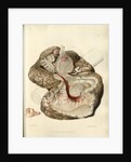
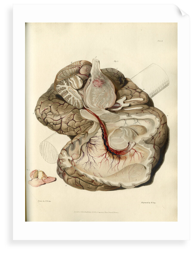
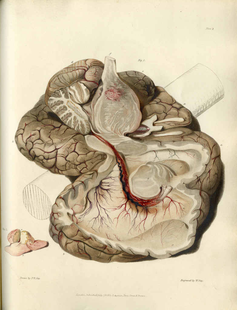
A section has been made through the pons Varolii; the left lateral ventricle having been drawn open to display its extent. The image is annotated: a. The anterior lobes of the cerebrum b. The posterior lobes of the cerebrum c. The middle lobe of the right hemisphere d. Part of the cerebellum e. The section of the cerebellum f. The commencement of the spinal cord g. The section of the pons Varolii showing a general enlargement of its substance by a gelatinous infiltration amongst its fibres h. The olfactory nerves i. The optic nerves j. The infundibulum distended with serum k. The choroid plexus l. The superior part of the lateral ventricle m. The posterior cornu of the lateral ventricle n. The anterior cornu of the lateral ventricle o. The brain thrown back, which if replaced would correspond with l p. The corpus striatum q. The optic thalamus Fig.2 û small auxiliary image. A scrofulous tumour. Plate 3 from Bright's Medical Reports: Diseases of the brain and Nervous System part 1 by Richard Bright (London, Longman, Rees... 1831). Inscribed: 'Plate 3. Drawn by F. R. Say. London Published July 1st. 1829. by Longman, Rees, Orme & Brown. Engraved by W. Say'
Original: engraving. 1831
- Image reference: RS-10323
- The Royal Society
Discover more
More by the artist William Say.
Explore the collection Science and anatomy prints and drawings.
Our prints
We use a 240gsm fine art paper and premium branded inks to create the perfect reproduction.
Our expertise and use of high-quality materials means that our print colours are independently verified to last between 100 and 200 years.
Read more about our fine art prints.
Manufactured in the UK
All products are printed in the UK, using the latest digital presses and a giclée printmaking process.
We only use premium branded inks, and colours are independently verified to last between 100 and 200 years.
Delivery and returns
We print everything to order so delivery times may vary but all unframed prints are despatched within 2-4 days via courier or recorded mail.
Delivery to the UK is £5 for an unframed print of any size.
We will happily replace your order if everything isn’t 100% perfect.