'Congestion in the minute vessels of the brain' William Thomas Fry

Product images of 'Congestion in the minute vessels of the brain'


Sizing information
| Overall size (inc frame) | x cm ( x in) |
| Depth | cm (in) |
| Artwork | x cm ( x in) |
| Border (mount) |
cm
top/bottom
(in)
cm left/right (in) |
| The paper size of our wall art shipped from the US is sized to the nearest inch. | |
 zoom
zoom
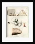
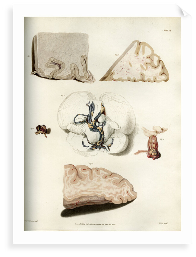
'Congestion in the minute vessels of the brain'
Illustration of multiple parts of the brain showing congestion in the vessels. Fig. 1- top left hand image. A portion of the brain when seen through an ordinary lens. Bright states; 'The whole medullary substance was found to be covered by fine grey specks and short hair-like vessels, resembling the appearance produced by scraping the nap of fine cloth over a sheet of white paper.' Fig.2- central image. Ossification of the vessels at the base of the brain. The under part of the cerebellum, the pons Varolii and the medulla oblongata are depicted in outline. The vertebral and basilar arteries proceeding from them are in a highly diseased state. This condition is common to old age and is frequently found in those who have died of apoplexy (hemorrhage/ stroke). Fig.3 - central left image. Example of an aneurism. Fig.4 - central right image. Disease of the choroid plexus. Fig.5- bottom image. The marbling apparent in the brain when congestion has taken place; this image was taken from a man who died of bronchitis. Fig.6 û top right image. The marbling apparent in the brain when congestion has taken place; the drawing was taken from a man who also had lungs that were extensively hepatized and oedematous (swollen from excessive fluid). Plate 19 from Bright's Medical Reports: Diseases of the brain and Nervous System part 2 by Richard Bright (London, Longman, Rees... 1831). Inscribed: 'Plate 19. F. .R. Say et C. J. Canton. Delt. London Published July 1. 1829. by Longman, Rees, Orme and Brown. W. T. Fry. Sculp.
Original: engraving. 1831
- Image reference: RS-10314
- The Royal Society
Discover more
More by the artist William Thomas Fry.
Explore the collection Science and anatomy prints and drawings.
Our prints
We use a 240gsm fine art paper and premium branded inks to create the perfect reproduction.
Our expertise and use of high-quality materials means that our print colours are independently verified to last between 100 and 200 years.
Read more about our fine art prints.
Manufactured in the UK
All products are printed in the UK, using the latest digital presses and a giclée printmaking process.
We only use premium branded inks, and colours are independently verified to last between 100 and 200 years.
Delivery and returns
We print everything to order so delivery times may vary but all unframed prints are despatched within 2-4 days via courier or recorded mail.
Delivery to the UK is £5 for an unframed print of any size.
We will happily replace your order if everything isn’t 100% perfect.